
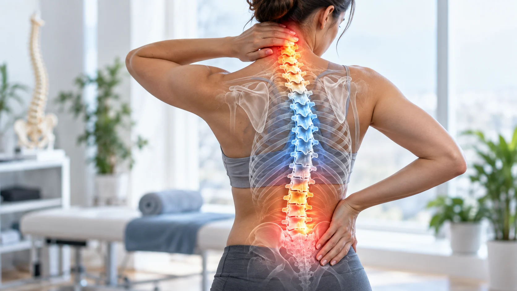
What Are Vertebrae?
Vertebrae are the individual bones that stack together to form the spinal column, commonly called the spine or backbone. These bones support the body, protect the spinal cord, and provide attachment points for muscles and ligaments. Although the spine may look like one continuous structure, it is made up of multiple vertebrae arranged in distinct regions.
A typical vertebra has a thick front portion called the vertebral body and a bony arch behind it. Together, these structures surround an opening through which the spinal cord travels. Additional projections, known as processes, provide places where muscles and ligaments attach and where neighboring vertebrae form joints.
The shape of each vertebra changes slightly depending on its location and function. Vertebrae in the neck are relatively small and mobile, while those in the lower back are larger because they carry more body weight. Understanding these differences makes a labeled spine much easier to interpret.
How Are the Vertebrae Labeled?
Vertebrae are labeled using letters and numbers based on their location in the spine. The cervical vertebrae are labeled C1 through C7, thoracic vertebrae T1 through T12, and lumbar vertebrae L1 through L5. Below these are the sacrum and coccyx, which contain vertebrae that become fused together.
The number generally increases as you move downward. For example, C1 is located at the very top of the cervical spine, while C7 sits at the bottom of the neck. T1 begins the thoracic region, and L5 is the lowest individual lumbar vertebra before the sacrum begins.
These labels help healthcare professionals identify exact locations when discussing injuries, imaging, arthritis, disc problems, or nerve symptoms. Instead of simply saying “lower back,” a medical report may describe a specific level such as L4-L5. Learning the labeling system can therefore make medical information easier to understand.
Cervical Vertebrae: C1 to C7
The cervical spine contains seven vertebrae located in the neck. These bones are labeled C1 through C7 and support the head while allowing a wide range of movement. They help you look upward and downward, rotate your head, and tilt it from side to side during everyday activities.
C1 is called the atlas because it supports the skull, while C2 is called the axis because it helps the head rotate. These first two cervical vertebrae have shapes that differ from typical vertebrae. Their specialized structure allows the upper neck to provide both stability and considerable mobility.
The lower cervical vertebrae, C3 through C7, look more similar to one another and help support the neck during movement. Nerves leaving this region contribute to sensation and muscle control in the shoulders, arms, and hands. Problems in the cervical spine can therefore sometimes cause symptoms beyond the neck itself.
Thoracic Vertebrae: T1 to T12
The thoracic spine contains twelve vertebrae labeled T1 through T12. This region begins below the cervical spine and extends through the upper and middle back. Each thoracic vertebra connects with one or more ribs, which makes this area important for supporting the rib cage and protecting organs in the chest.
Thoracic vertebrae allow rotation and some bending, but the rib cage limits their movement compared with the neck and lower back. This relative stiffness provides stability for the trunk. It also creates a strong framework for muscles involved in posture, breathing, shoulder movement, and upper-body control.
Upper-body exercises can place controlled demands on this region. Movements such as the seated chest press rely on a stable upper back and trunk while the arms push forward. This does not mean the thoracic vertebrae produce the pressing force, but they provide structural support during the exercise.
Lumbar Vertebrae: L1 to L5
The lumbar spine contains five vertebrae labeled L1 through L5. These bones are located in the lower back between the thoracic spine and sacrum. Lumbar vertebrae have relatively large vertebral bodies because they must support substantial body weight during standing, walking, lifting, and many other activities.
The lumbar region allows flexion, extension, and some side bending, while rotation is more limited. This combination of mobility and load-bearing ability makes the lower back important for everyday movement. Muscles surrounding the lumbar spine, abdomen, hips, and pelvis work together to control these forces.
L4-L5 and L5-S1 are commonly discussed spinal levels because they experience considerable mechanical loading. Pain in this region does not automatically mean a disc or vertebra is damaged, however. Lower-back symptoms can involve muscles, joints, nerves, discs, or several factors working together.
The Sacrum and Coccyx
The sacrum sits below the lumbar spine and forms the back portion of the pelvis. It is made from five vertebrae, usually labeled S1 through S5, that fuse together during development. The sacrum connects the spine with the pelvis and helps transfer forces between the upper body and legs.
On either side, the sacrum connects to the pelvic bones through the sacroiliac joints. These joints allow only small amounts of movement but play an important role in force transfer during walking, standing, and lifting. Muscles and ligaments around the pelvis provide additional support to this region.
Below the sacrum is the coccyx, commonly called the tailbone. It is generally made up of several small fused vertebrae, although the exact number can vary. The coccyx provides attachment points for pelvic floor muscles and ligaments and can become painful after falls or prolonged pressure.
What Is the Main Function of the Vertebrae?
One of the primary functions of vertebrae is protecting the spinal cord. The openings within individual vertebrae align to create the spinal canal, forming a protective bony tunnel. The spinal cord carries messages between the brain and much of the body, making this protective function extremely important.
Vertebrae also support body weight and help maintain the overall structure of the trunk. The lower spinal bones carry greater loads than those near the top, which is why lumbar vertebral bodies are larger. Their stacked arrangement allows the spine to remain strong while still providing useful movement.
Another function is providing attachment points for muscles, tendons, and ligaments. These tissues work together to move and stabilize the spine during bending, lifting, walking, and exercise. Without these attachment points, the body would have far less control over posture and trunk movement.
How Intervertebral Discs Work With Vertebrae
Intervertebral discs sit between most adjacent vertebral bodies and help absorb and distribute forces through the spine. Each disc has a softer central region surrounded by a stronger outer layer. This structure allows the disc to deform slightly as the spine moves and loads change throughout the day.
Discs also create space between vertebrae, helping maintain room for spinal nerves to exit. They contribute to flexibility by allowing small movements between individual spinal segments. Combined across many spinal levels, these small motions allow the spine to bend, extend, and rotate through useful ranges.
Discs naturally change with age, and imaging often shows changes even in people without pain. A disc finding therefore does not automatically explain symptoms. Healthcare professionals usually interpret imaging alongside a physical examination, medical history, and the specific pattern of pain or neurological signs.
Facet Joints and Spinal Movement
Facet joints are small joints located at the back of the vertebrae. Each vertebra forms facet joints with the bones above and below it, helping guide spinal movement while providing stability. Their orientation differs between spinal regions, which contributes to differences in mobility between the neck, mid-back, and lower back.
In the cervical spine, facet joints allow considerable movement, supporting rotation and bending of the head. Thoracic facets work alongside the rib cage to create more controlled rotation, while lumbar facets favor flexion and extension. This regional design helps each section of the spine perform its specific role.
Facet joints can become irritated due to injury, arthritis, repetitive loading, or other changes. Symptoms may include localized back or neck discomfort and stiffness. Because similar symptoms can come from muscles, discs, or other structures, professional assessment may be needed when pain is persistent or significantly limits movement.
Common Problems Affecting the Vertebrae
Fractures can affect vertebrae after significant trauma, falls, or conditions that weaken bone. Compression fractures are more common in people with reduced bone density and may cause sudden back pain or changes in posture. Significant trauma or sudden severe spinal pain requires medical evaluation rather than relying solely on home exercises.
Arthritic changes can develop around the joints between vertebrae as people age. These changes may be visible on imaging without causing symptoms, while some people develop stiffness or discomfort. Treatment decisions are usually based more on symptoms and function than on imaging findings alone.
Structural changes such as scoliosis, spondylolisthesis, or age-related degeneration can also affect the spinal column. These terms describe different conditions and should not be treated as interchangeable. Some people have noticeable spinal changes with few symptoms, while others require rehabilitation, monitoring, or medical treatment depending on severity.
Nerves, Vertebrae, and Radiating Pain
Spinal nerves leave the spinal canal through openings between neighboring vertebrae. These nerves carry signals controlling sensation and movement throughout the body. If a nerve becomes irritated or compressed, symptoms may appear away from the spine in an arm, hand, leg, or foot depending on the affected level.
Cervical nerve irritation can sometimes produce pain, tingling, numbness, or weakness in the upper limb. Problems involving lumbar nerve roots may create symptoms traveling into the buttock or leg. The exact pattern can help healthcare professionals identify which nerve structures may need further assessment.
Radiating pain does not always mean severe nerve damage. Nerves can become temporarily sensitive for several reasons, and many symptoms improve with appropriate care. Progressive weakness, major numbness, or changes in bladder or bowel function are more concerning and require prompt medical evaluation.
How to Support a Healthy Spine
Regular physical activity is one of the simplest ways to support spinal health. Walking, strength training, and recreational movement help maintain muscle capacity and general mobility. The spine is designed to tolerate movement, so avoiding activity completely is rarely the best long-term strategy for otherwise healthy people.
Strengthening the trunk, hips, and upper back can improve the body’s ability to manage everyday loads. Exercises such as rows, squats, carries, bridges, and controlled core movements can all contribute. There is no single exercise that guarantees a healthy spine, so a balanced routine is usually more practical.
Daily habits also matter, particularly when work involves long periods of sitting or repetitive tasks. Change position regularly, adjust your workspace for comfort, and avoid relying on one supposedly perfect posture. Adequate sleep, gradual training progression, and sensible lifting habits can further support comfortable movement.
When to Get Medical Help for Spine Symptoms
Seek medical attention after significant trauma if you develop severe neck or back pain, weakness, or difficulty moving normally. Falls, vehicle accidents, or major sports collisions can injure bones, discs, ligaments, or nerves. A healthcare professional can determine whether imaging or other tests are appropriate.
Persistent pain should also be evaluated when it continues for weeks, repeatedly worsens, or interferes with sleep and everyday activity. Back and neck pain have many possible causes, and vertebral problems are only one category. A proper examination can help identify whether rehabilitation, medication, further testing, or another approach is appropriate.
Urgent medical care is important for new bladder or bowel control problems, numbness around the groin, rapidly progressing weakness, or severe neurological symptoms. Fever with severe spinal pain or major unexplained symptoms also deserves prompt assessment. These warning signs should not be managed only with stretching or exercise at home.
Conclusion
Vertebrae are the bones that form the spinal column, protect the spinal cord, support body weight, and provide attachment points for muscles and ligaments. They are labeled according to region, including C1-C7 in the neck, T1-T12 in the thoracic spine, and L1-L5 in the lower back.
Below the lumbar region are the fused vertebrae of the sacrum and the smaller bones of the coccyx. Each spinal region has a different shape and movement role. Intervertebral discs, facet joints, muscles, and ligaments work alongside the vertebrae to create a spine that is both stable and mobile.
Many spinal changes are manageable and do not automatically indicate a serious problem. However, severe trauma, progressive weakness, significant numbness, or bladder and bowel changes require medical attention. Understanding vertebral labels can make medical discussions and spine-related information much easier to follow.
FAQs
How many vertebrae are in the human spine?
The spine typically begins with 33 vertebrae, although several fuse during development. Adults usually have 24 movable vertebrae, plus the fused sacral and coccygeal vertebrae.
What do C, T, and L mean in vertebrae labels?
C refers to cervical vertebrae, T refers to thoracic vertebrae, and L refers to lumbar vertebrae. The number shows the vertebra’s position within that particular spinal region.
What is the largest vertebra in the spine?
Lumbar vertebrae are generally the largest individual movable vertebrae because they support substantial body weight. Their vertebral bodies become relatively large compared with bones higher in the spine.
What is the difference between vertebra and vertebrae?
“Vertebra” refers to one individual spinal bone, while “vertebrae” is the plural term for two or more of these bones. The spinal column contains multiple vertebrae stacked together.
Can damaged vertebrae heal?
Some vertebral injuries, including certain fractures, can heal with appropriate treatment and time. Recovery depends on injury type, bone health, stability, neurological involvement, and the treatment recommended by a healthcare professional.